Planarian regeneration
Bogdanova E, Vagner L, Lukyanov S, Shagin D.
Owing to their extraordinary ability for regeneration and asexual reproduction, planaria have long been the model organisms for studying cellular and molecular mechanisms leading to morphological plasticity and pattern reestablishment. Un-like the other models for studying morphogenesis (insects, nematodes, fish, amphibians, and mammals; hydra is the only exception) turbellarians have no definite stage in their life cycle to which the major events of body plan formation are restricted. Instead, planarian is always in the process of remaking itself through continuous cell turnover, growing and shrinking in volume and length depending on external conditions, being always ready to restore itself even from a tiny fragment of its body. It can be said that, unlike most other animal phyla, the body patterning mechanism of planarians is always active while the animal is alive. Still, its molecular basis remains unknown.
Identification and characterization of a new family of C-type lectin-like genes from planarian
A novel family of C-type lectin-like genes, denoted multidomain free lectin (MDFL), was identified in the freshwater planaria Girardia (Dugesia) tigrina. We cloned several genes that encode proteins comprising a signal peptide and a number of consecutive C-type lectin-like domains (CTLDs) interconnected by short linker stretches. Full-length cDNAs and genomic sequences were obtained using Step-Out RACE, genome walking and PCR with gene specific primers. Analyses of genomic organization, CTLD amino acid sequences, and the overall architecture of these proteins indicate that planarian genes are a separate family of C-type lectin-like genes. Using whole-mount in situ hybridization, we demonstrated that MDFL genes are differentially expressed along the anterior-posterior axis of planarian body. Whole-mount immunostaining and electronic microscopy revealed that at least two MDFL proteins (Scarf2 and gtLec1) are produced by specifically differentiated gland cells with elongated cell processes and are excreted as components of the planarian body surface mucus. On the base of the analysis of literature data, we concluded that these gland cells are viscid glands of duo-type.
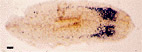
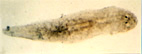
To examine the idea that the presence of body edge plays a role in support of gland excretion function we used the following model: The planarian heads was transplanted to the neck region of planarian body and two types of monsters were collected after 30 days of regeneration. First type does not have a body edge in ectopic part that was covered by the dorsal epithelium only. The second contains an ectopic body edge where dorsal and ventral epithelium interacted. Cell necks with positive immunostaining for gtLec1 were observed in the ectopic heads of the second type and was not found in the first type of heads.
Inductive interactions regulating body patterning in planarian, revealed by analysis of expression of scarf2 gene
We performed a series of experiments using one of the MDFL genes, scarf2 (previously scarf), as a molecular marker. Its expression was monitored during different types of regeneration by whole-mount in situ hybridization and RT-PCR. The obtained data suggest that scarf2 expression is positively regulated by anterior tissues closely adjacent to scarf2 expressing region, so that their surgical removal results in rapid scarf2 switch-off. In turn, tissues expressing scarf2 seem to inhibit its activation anteriorly. The dynamics and pattern of scarf2 expression variations during regeneration indicate that the body patterning in planarians is based on a system of hierarchial inductive interactions rather than on a global morphogen gradient.
Novel Planarian Extrachromosomal Virus-like Element identified by planarian genomic DNA subtraction
Despite extensive information on the biology and genetics of planaria, the occurrence and distribution of viruses in these animals remains an unexplored area of research. The planarian species (Planariidae family) are represented by different strains employing various modes of reproduction, including exclusively sexual, exclusively asexual, or both sexual and asexual. Using a combination of Suppression Subtractive Hybridization (SSH) and Mirror Orientation Selection (MOS), we compared the genomes of two strains (sexual and asexual) of freshwater planarian, Girardia tigrina.
The novel extrachromosomal DNA-containing virus-like element denoted PEVE (Planarian Extrachromosomal Virus-like Element) was identified in the asexual planarian strain. PEVE is the first viral element identified in free-living flatworms. The PEVE genome (about 7.5 kb) consists of two unique regions (Ul and Us) flanked by inverted repeats. PEVE differs from all known viruses and viral elements. Sequence analyses reveal that PEVE comprises two helicase-like sequences in the genome, of which the first is a homolog of a circoviral replication initiator protein (Rep), and the second is similar to the papillomavirus E1 helicase domain.
PEVE genome exists in at least two variant forms with different arrangements of single-stranded and double-stranded DNA stretches that correspond to the Us and Ul regions. PEVE is unevenly distributed and expressed in the worm body, and is detected in specific parenchyma cells.