
Подразделение было расформировано в 2024 году.
Группа синтетической биологии
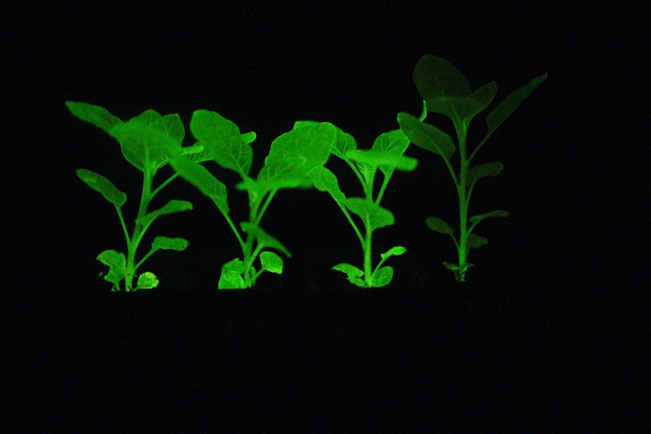
Группа Синтетической биологии сформирована в 2017 году, как часть Отдела биомолекулярной химии. Основной научной задачей группы является создание и развитие технологий биолюминесцентного имаджинга, с фокусом на неинвазивный имаджинг растений.
Помимо этого, в группе ведутся работы по белковому дизайну, расшифровке новых биолюминесцентных систем и определению генов биосинтеза природных соединений.
| ФИО | Должность | Контакты |
|---|---|---|
Ранее здесь работали | ||
| Балакирева Анастасия Васильевна, к.б.н. | ||
| Ветринская В.В. | ||
| Волков П.В. | ||
| Горбачев Дмитрий Андреевич, к.б.н. | ||
| Попова В.В. | ||
| Строкач Никита Николаевич | ||
| Дюф А.А. | ||
| Болотина В.С. | ||
| Чернышёва А.Н. | ||
 Загрузка...
Научные проекты
Загрузка...Москва, ул. Миклухо-Маклая, 16/10 — На карте
Загрузка...