
Glowing fungi expose final enzyme that could make bioluminescent tools more efficient
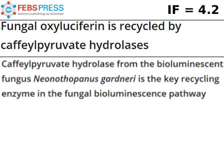
Researchers from the Department of Biomolecular Chemistry at the Shemyakin-Ovchinnikov Institute of Bioorganic Chemistry as part of an international collaboration, have published two papers in The FEBS Journal confirming the role of the CPH enzyme in the fungal bioluminescence pathway. The team showed that this enzyme breaks down oxyluciferin into caffeic and pyruvic acids. Caffeic acid then returns to the bioluminescent system and helps sustain light emission. This metabolite recycling mechanism helps explain how fungi maintain bioluminescence and may reduce the energetic cost of the process. The findings open up new opportunities for developing more efficient autonomous bioluminescent systems, with potential applications in medicine, biotechnology, agriculture, and environmental monitoring. Learn more
News ")
- The stepwise mechanism of TRPV6 channel blockade by polyamine spermine
science news
VI.3 Researchers from IBCh RAS, as part of an international collaboration, have elucidated the mechanism by which the natural polyamine spermine blocks the human calcium channel TRPV6. The study was supported by the Russian Ministry of Science and Higher Education (computational modeling) and published in Nature Communications.
- Glowing fungi expose final enzyme that could make bioluminescent tools more efficient
science news
V.22 Researchers from the Department of Biomolecular Chemistry at the Shemyakin- Ovchinnikov Institute of Bioorganic Chemistry together with international collaborators published two papers in The FEBS Journal confirming the role of the CPH enzyme in the fungal bioluminescence pathway: it breaks down oxyluciferin into caffeic and pyruvic acids, helping sustain light emission by recycling caffeic acid back into the bioluminescent system, while pyruvic acid may be redirected into central metabolism to help generate cellular energy. The findings open up opportunities for developing more efficient autonomous bioluminescent systems for medicine, biotechnology, agriculture, and environmental monitoring.
- How can plant defence responses to pathogen and pest attacks be observed?
science news
III.18 Scientists from the Department of Biomolecular Chemistry at the Shemyakin–Ovchinnikov Institute of Bioorganic Chemistry of the Russian Academy of Sciences developed autoluminescent reporters for non-invasive imaging of plant defence responses. Using consumer-grade cameras, they visualized the spatiotemporal activity of the phytohormones salicylic acid and jasmonic acid in Arabidopsis thaliana and Nicotiana benthamiana during normal development as well as in response to pest and pathogen attacks. The study was published in Nature Communications.
Events
- Open seminar of the Department of Immunology
science news
VI.8 (This event is over) Dear colleagues! On June 8 (Monday) at 3 p.m., an open seminar of the Department of Immunology will be held in seminary room 404 (4th floor, 52nd building), dedicated to the pre-defense of the dissertation for the degree of Candidate of Biological Sciences Anastasia I. Palamarchuk on the topic: "Overexpression of the telomerase catalytic subunit gene as a factor influencing the transcriptional and functional activity of NK cells, as well as their sensitivity to targeted elimination", specialty 1.5.3. Molecular Biology. Everyone is invited to attend. June 8th
- Symposium under the auspices of the Russian Neurochemical Society, dedicated to Eugene Grishin’s 80th anniversary
science news
IV.20 (This event is over) The symposium, organized by the Department of Molecular Neurobiology, will take place on April 20 at 11:00. The program includes the unveiling of a memorial booth and presentations by colleagues, students, friends, and associates of Eugene Grishin. We cordially invite everyone interested in modern research in the field of toxins, ion channels, and neurobiology! The presentations will be broadcast via Zoom.