
Press-room / news / Science news /
Why is the bush cricket green?
A team of scientists with the participation of researchers from the Institute of Bioorganic Chemistry RAS has revealed the nature of the green pigment in the bush cricket Tettigonia cantans. It turned out that the bush cricket owes its protective coloration to a chromoprotein with a unique fold that contains two chromophores simultaneously. One of them is a yellow carotenoid, and the other is a blue bilin. The mixture of the two colors results in a bright green coloration, practically indistinguishable from grass and allowing the animal to cleverly hide. The work has been published in PNAS.

The molecular nature of green insect coloration has been a subject of debate since at least the late 19th century. Our work has finally provided an answer. Using extraction and several stages of chromatography and precipitation, a protein pigment called DBXN was purified from the green integument of bush crickets. The spatial structure of DBXN was studied using X-ray crystallography, and a new type of fold was discovered. Two identical protomers (≈40 kDa) consist of three polypeptide fragments that are formed from a large precursor protein as a result of post-translational proteolysis. A large hydrophobic cavity simultaneously contains two molecules of carotenoid (lutein) and bilin (hydroxyethylfarnesylated mesobiliverdin) and four molecules of phosphatidylcholine (see Figure). Preliminary data show that the green coloration of other insects may be the result of different combinations of pigments, in some cases a similar mechanism is realized when two different chromophores are bound to the same protein. The work was led by Nikolai Sluchanko from the A.N. Bach Institute of Biochemistry, Russian Academy of Sciences, and Rustam Ziganshin and Alexander Vassilevski from the Institute of Bioorganic Chemistry RAS participated in it.
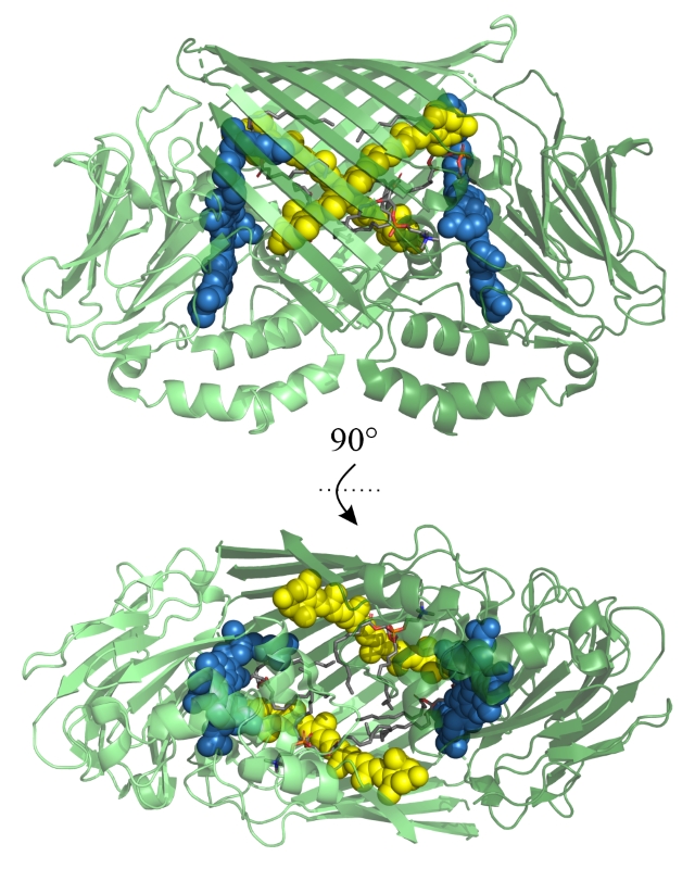
Figure. Spatial structure of DBXN (PDB ID: 9KUE). Polypeptide chains are shown in ribbon
representation, chromophore atoms are shown as spheres, chemical bonds in lipid molecules are shown
as sticks. Two protein protomers are colored light and dark green, carotenoid molecules are yellow, bilin
is blue, and phosphatidylcholine tails are gray.
may 30, 2025