
Group of electron microscopy
Electron and scanning probe microscopy methods have already become routine instruments in laboratory research. Currently, the main interest of scientists in this field is focused on the continuous development of methods for samples deposition, preparing substrates and probes for improvement of quality and reproducibility of the results, expanding the scope of applicability of microscopy methods.
Thus, the development of super-sharp probes for atomic force microscopy made it possible to visualize the details of DNA molecules and proteins and their constituent parts. New methods for the preparation of substrates made it possible to apply biological objects without changing their structure to obtain their true image. Various laboratories are conducting research aimed at using films and nano-thickness grids as substrates for electron microscopy. This will increase the contrast and information content of images, avoid contrasting.
In our group, we concentrate our efforts on the development of new approaches in electron and atomic force microscopy of biological objects using nanostructured materials.
During several years the group of electron microscopy research was aimed at a more effective use of the atomic force microscopy method for visualizing biomacromolecules. Previously, we have empirically determined the conditions for obtaining nanostructures at the tips of standard atomic force microscopy probes, which made it possible to improve the resolution of the method by about an order of magnitude (from 15–20 nm to ~ 2 nm). In 2018, we completed a study of the physical properties of these structures, explaining the growth mechanisms and efficiency in microscopy. Based on the results of work, an article was published in the journal Ultramicroscopy, which is part of 1 quartile. In 2017, 2 reports were presented at conferences.
The synthesis takes place using the method of gas-phase chemical deposition activated by plasma. Statistical analysis of electron microscopy images shows that the growth rate of branched nanostructured spikes is of the order of 100 nm / h. The radius of curvature of the resulting structures is 1-2 nm. The results of a study of the obtained probes by Raman scattering showed that the branched structures consist of amorphous carbon and, likely, glassy carbon, taking into account high mechanical characteristics.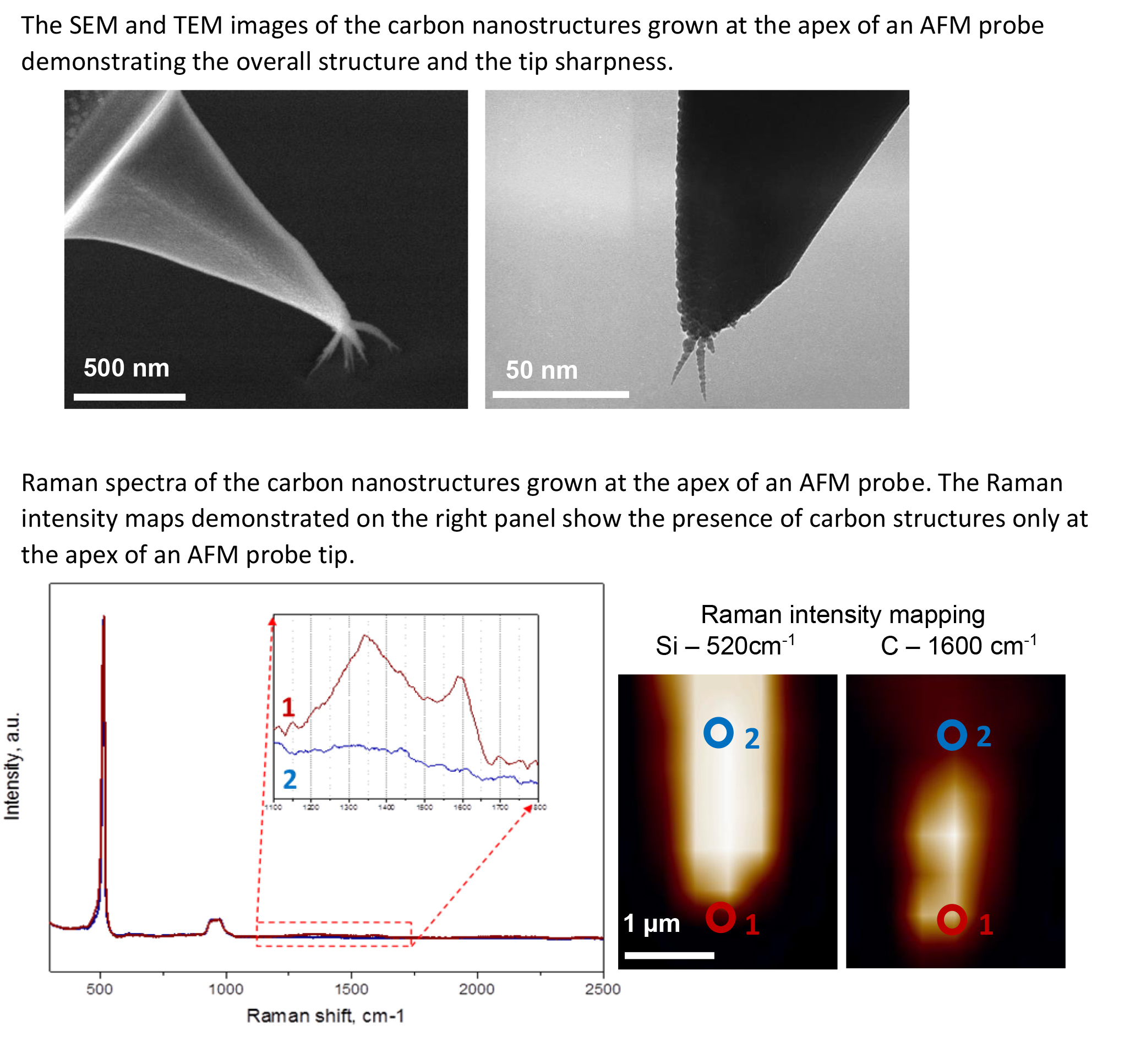
| Fullname | Position | Contacts |
|---|---|---|
Previously worked here | ||
| Ekaterina Obraztsova, Ph.D. | ||
| Pavlova E.R. | ||
| Obraztsova A.S. | ||
| Men'chish R.M. | ||
| Tuyakova F.T. | ||
 Loading...
Scientific projects
Loading...Ekaterina Obraztsova
Russia, Moscow, Ul. Miklukho-Maklaya 16/10 — On the map
Loading...