
Press-room / news / Science news /
Scientists made proteins shine longer
Russian scientists together with foreign colleagues extended the shine time of fluorescent label in live cells. Light is emitted longer due to the continuous exchange of fluorogenic dyes interacting with special protein labels. The method which was described in the Chemical Science journal will simplify the live cells staining and provide new opportunities for research. This work was supported by the Russian Science Foundation.
The ability to absorb light at short wavelengths and re-emit it at longer wavelengths is called fluorescence. Biophysicists, molecular and cellular biologists are using this physical phenomenon in their work. Scientists label proteins to see their location and interaction inside live cell. Green fluorescent proteins (GFP) from the jellyfish Aequorea victoria and their analogues of other colors are often used for this. They are genetically encoded tags that fluoresce by themselves, without the chemicals addition from outside. Another way is to mark the proteins with labels irreversibly binding the light-emitting molecules (chemical fluorophores). This process allows choosing the spectra of light absorption, fluorescence and labeling time. But the method requires long preparation and washing out of the excess dye.
“Many fluorophores penetrate through the live cell membrane badly and bind to protein-partners slowly,” explains Alexander Mishin, researcher from the IBCh RAS, Laboratory of Biophotonics. “To speed up the staining, fluorophores are added in excess. Therefore, free fluorophores accumulate in the cell. They create an unwanted background glow, in which noise "overlaps" useful signals. Therefore, one has to wash out the cells before image acquisition. It increases the staining time, and therefore such labels are not suitable for studying fast processes. For example, to study cytoskeleton, a dynamic system inside the cell.”
Fluorogenic dyes, which start to work not in solvents, but in combination with a certain molecule, help to avoid this.
In the new study, researchers from the Institute of Bioorganic Chemistry of the Russian Academy of Sciences, the Nizhny Novgorod State Medical Academy and Pirogov Russian National Research Medical University together with their Spanish colleagues from the Center for Genomic Regulation created "fluorogen-protein" pairs for protein labeling in live cells. Such "duets" help the scientists to extend the glow time. Light is emitted longer due to continuous exchange of fluorogenic dyes, which interact with genetically encoded tags. Selected fluorogens penetrate into live cells and color the "target" quickly. The developed "fluorogen-protein" pairs are well-suited in super resolution fluorescence microscopy, based on fluorescent dyes’ molecules “outbursts”.
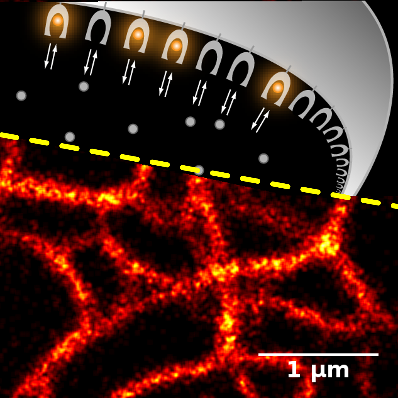
Above: molecules of fluorogen, emitting light molecules (gray and orange circles), begin to glow in combination with a special protein (genetically coded label). The constant replacement of spoiled fluorogen molecules with new ones protects against burnout under the influence of strong light. The moment of combining the molecule and protein is seen in the microscope as a flash. Below: analysis of many such flares can be used to reconstruct an ultrahigh resolution image (here - part of the cytoskeleton).
“We used such methods as computer simulation of "fluorogen-protein" interaction and chemical synthesis of fluorogens library when developing a new tagging system,” Alexander continues. “Many types of microscopy are inaccessible in Russia. But we solved this problem with the help to foreign colleagues participation in our study”.
According to the authors, the new method based on fluorogenic dyes will simplify the cells staining greatly and allow scientists to use a more complex experimental design in their studies.
september 1, 2017